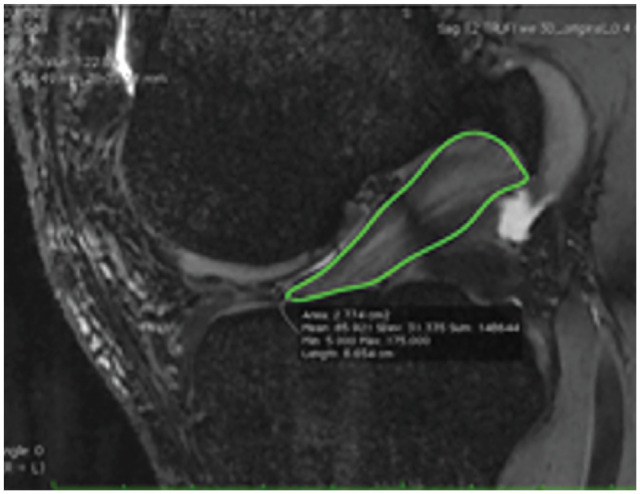
Figure 3.

Mapping involved manually drawing a region of interest around the ACL on each image where the ACL was visible on the 3D true-FISP series of each MRI. ACL, anterior cruciate ligament; FISP, fast imaging with steady-state precession; MRI, magnetic resonance imaging; 3D, 3-dimensional.
