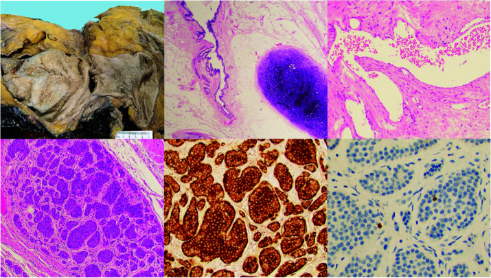
Figure 3.
Top left: Gross photograph of the tumor demonstrates predominantly solid composition with large cystic areas (5 cm ruler for scale). Top middle: H&E stain showing a mature cystic teratoma with epithelial and mesenchymal elements. Top right: 40× H&E stain showing a focus of erectile-type vascular tissue. Bottom: 20× H&E showing incidental focus of well differentiated neuroendocrine tumors with diffuse expression of chromogranin and a low Ki67 proliferation index.

