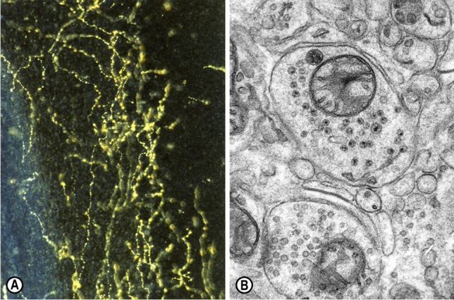
Fig. 1.

(A) Serotonin-immunoreactive fibers in the mouse cerebellum, displaying their typical axonal varicosities. Sternberger peroxidase-antiperoxidase (PAP) method, dark-field illumination, × 40 [18]. (B) Electron micrograph of a monoaminergic varicosity or bouton en passant in the molecular layer of the cerebellum of a “Purkinje cell degeneration” (Agtpbp1pcd/Agtpbp1.pcd) mutant mouse, containing small granular vesicles (40–60 nm in diameter) and a large granular vesicle (90–110 nm in diameter). Potassium permanganate (KMnO4) fixation method, ultrathin section stained with uranyl acetate and lead citrate, × 24,000 [16]
