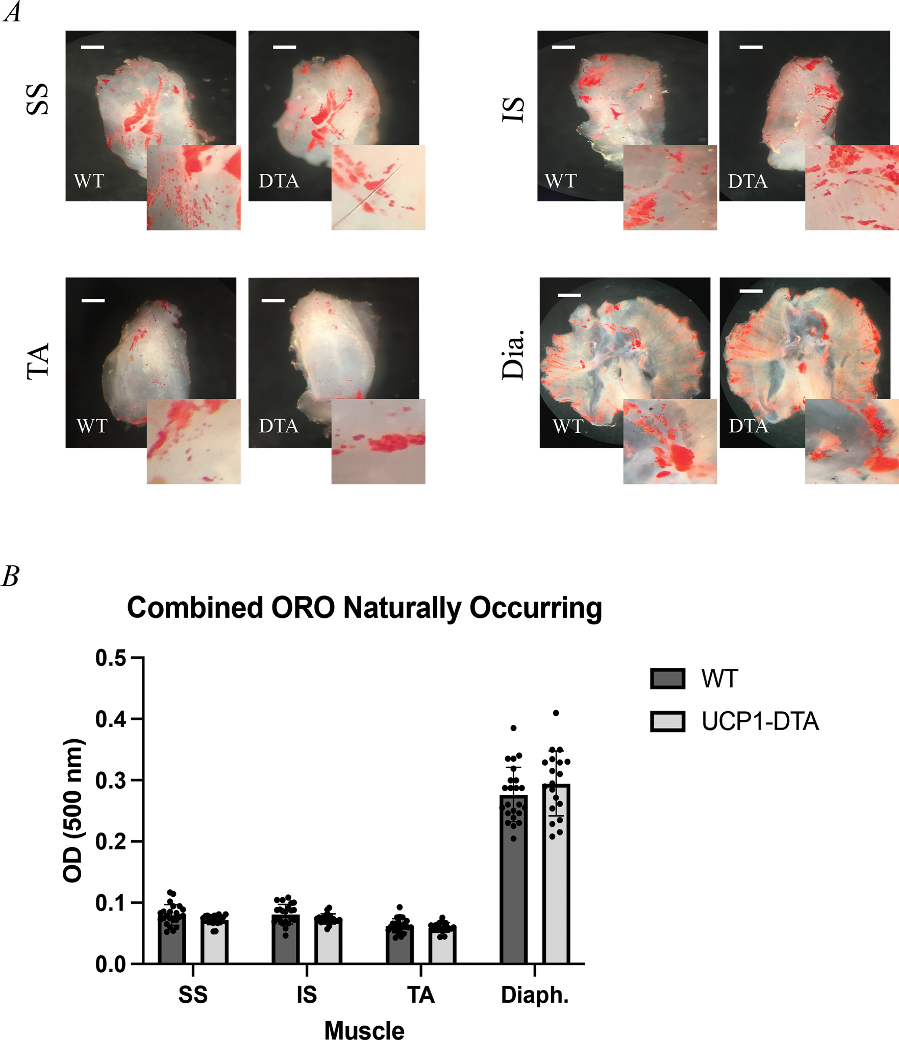
Figure 3. Intramuscular Adipose Tissue (IMAT) Develops Without UCP-1-Expressing Cells.

Oil Red O staining of decellularized A Supraspinatus (SS), Infraspinatus (IS), Tibialis Anterior (TA), and Diaphragm (Dia.) muscles depicting visually similar amounts of naturally occurring IMAT (red) in both wild type (WT) and UCP1-DTA genotypes. B Optical density (OD) measurement confirms similar amount of lipid content across the four muscles in both female (n=14) and male (n=13) mice. Scale bar 1mm. Analysis performed in Prism using multiple, independent t-tests. Statistical significance set at p<0.05.
