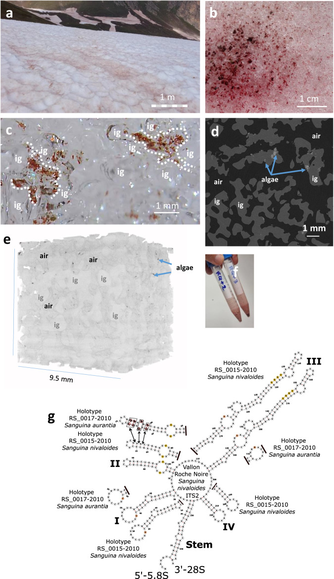
Fig. 1. Sanguina nivaloides blooms.
a Red snowfield in Vallon Roche Noire, 2300 m. a.s.l. The bar shows the scale in front of the perspective view. b Algal bloom view at the melting snow surface. c Red cysts in the liquid water fraction circulating between ice grains. The liquid water content, measured 10 cm below the snow surface, was 16 ± 4 mass %. Imaging with field digital microscope shows cysts only in the liquid water fraction, moving along water currents in interstices between ice grains. This observation was repeated 5 times with similar result. d X-Ray imaging of red snow. Air is shown in black, ice grains in dark gray, dust particles in white, and clusters of cysts in light grey (arrows) at the periphery of ice grains. e Volume view of red snow analyzed by X-Ray tomography. Cysts are detected at the periphery of ice grains, facing the reticulated air network. f Collected algal cells for laboratory analysis. Although present at the surface of snow, cysts sedimented after melting. g Assessment of algal species present in collected blooms based on ITS2 analysis. ITS2 secondary structures (including the ‘stem’ 5’− 5.8 S rRNA and 3’− 28 S rRNA) of algae sampled in Vallon Roche Noire and all other locations in this study, were predicted using RNAfold according to centroid algorithm. Obtained ITS2 structures were compared to S. nivaloides holotype RS 0015–2010 and S. aurantia holotype RS 0017–2010. All sequences matched holotype RS 0015-2010. Differences with holotype RS 0017–2010 are highlighted in pink, where additional complementary bases are present in helix II (5’-CG-3’, 5’-UA-3’, 5’-GA-3’). Single bases colored in brown are also different in helix I and III of S. aurantia in comparison to S. nivaloides. Pyrimidine - pyrimidine mismatch in Helix II and (G/U)GGU motif in helix III, specific of Viridiplantae, are shown in yellow. Black lines indicate no difference downstream of the structure. ig, ice grain.

