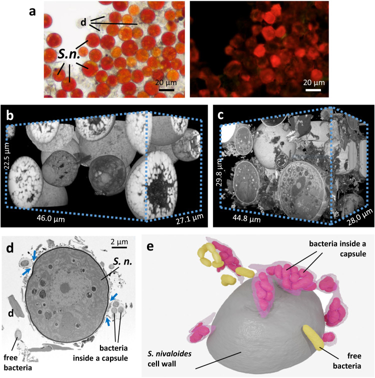
Fig. 3. FIB-SEM imaging of chemically-fixed and cryo-fixed S. nivaloides cysts.
a Light microphotographs of red snow. Right, bright field image of mature cysts, with lipids droplet visible inside cells, pigmented in red or orange due to the presence of astaxanthin and other carotenoids. Left, chlorophyll autofluorescence (excitation 495 nm; emission 521 nm). Fluorescence intensity is higher in orange cells and cells with apparent green spots. Debris and dust in the sample have no pigmentation nor fluorescence. This observation has been repeated 6 times with similar results. b Volume view of chemically-fixed cells analyzed by FIB-SEM. One FIB-SEM analysis has been performed with this fixation method. c Volume view of cryo-fixed cells analyzed by FIB-SEM. One FIB-SEM analysis has been performed with this fixation method. d Detection of bacteria associated to Sangina nivaloides cysts. Image from a FIB-SEM stack showing free and encapsulated bacteria. This observation has been repeated with similar result on all images from the FIB-SEM stack. e Three-dimensional model of bacteria at the vicinity of a Sangina nivaloides cyst. S. n. Sangina nivaloides, d debris.

