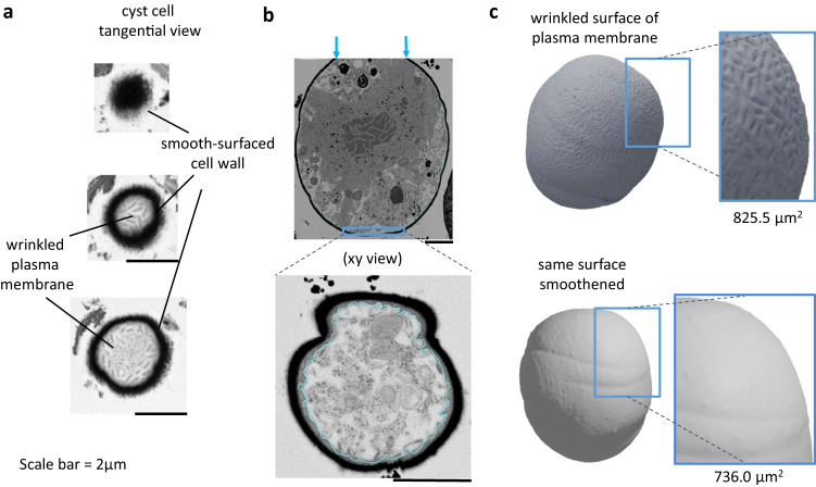
Fig. 4. Wrinkled plasma membrane of Sanguina nivaloides cysts.
a Tangential views of plasma membrane and cell wall in cross sections observed by FIB-SEM at variable depths. b Process of segmentation of plasma membrane based on EM image stacks. c Three-dimensional reconstruction of Sanguina nivaloides cyst plasma membrane. The 3D model with a wrinkled surface was filtered using the HC Laplacian smoothing method77. The filter builds a new mesh based on the information of the average of the nearest vertices, and thus produces a smooth surface after three iterations. Based on the native wrinkled membrane and the computed smoothened one, surfaces are calculated and highlight a 12% area increase attributable to the plasma membrane architecture.

