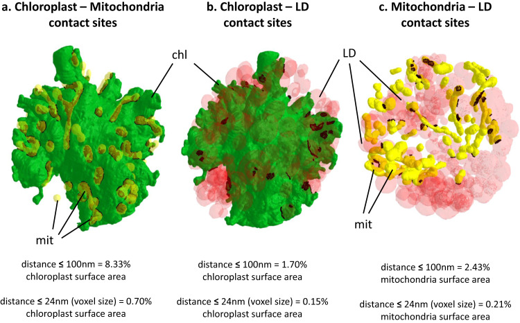
Fig. 8. Proximity between cellular organelles in S. nivaloides cysts.
a Chloroplast - mitochondria. b Chloroplast - lipid droplets. c Mitochondria – lipid droplets. Distances were measured as described earlier24. Green, plastid surface; yellow, mitochondria surface; red, lipid droplets surface. Dark spots highlight proximity surfaces corresponding to points at a distance ≤ 100 nm between organelles. chl chloroplast, LD lipid droplet, mit mitochondria.

