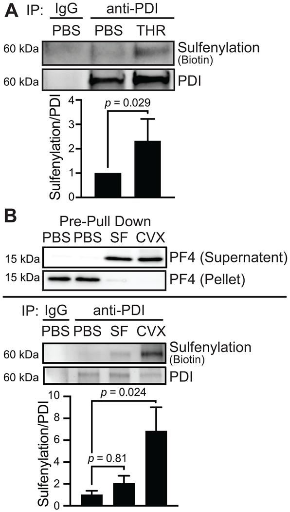
FIGURE 6.

Endothelial cells and platelets secrete sulfenylated PDI. (A) PDI secreted from activated endothelial cells is sulfenylated. HUVECs were stimulated with 1 U/mL human a thrombin (THR) in the presence of 0.1 mM BTD for 1 h before inactivation with 5 U/mL hirudin and media collection for immunoprecipitation. The media was concentrated before immunoprecipitating PDI with an anti-PDI or control IgG antibody. Click chemistry was performed to attach biotin-PEG3-azide onto the alkyne arm of BTD, and biotin was detected by western blotting and chemiluminescence. (B) PDI secreted from activated platelets is sulfenylated. Washed human platelets (1 × 10⁹/mL) were stimulated with 50 μM SFLLRN (SF) or 500 ng/mL convulxin (CVX) for 15 min in the presence of 0.1 mM BTD. The supernatant containing the releasate was collected after centrifuging the platelets, and a biotin-PEG3-azide was conjugated to the alkyne of BTD by click chemistry. The releasate was desalted, and PDI was immunoprecipitated with an anti-PDI or control IgG antibody. Biotin was detected by western blotting and chemiluminescence. A control blot to assess platelet factor 4 (PF4) as a marker of platelet secretion was performed in the releasate and the cell pellet before immunoprecipitation. p-values were determined by unpaired Student’s t-test (A) and one-way ANOVA with Dunnett’s post hoc analysis (B). Data represented as mean ± SEM.
