FIGURE 2.

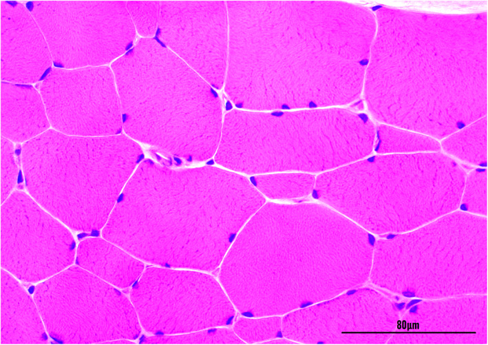
Transverse section of the biceps femoris muscle of cat 1 with hereditary myotonia showing variation in the muscle fiber diameter and hypertrophy. Hematoxylin and eosin stain (×400). Magnification bar = 80 μm.

Transverse section of the biceps femoris muscle of cat 1 with hereditary myotonia showing variation in the muscle fiber diameter and hypertrophy. Hematoxylin and eosin stain (×400). Magnification bar = 80 μm.