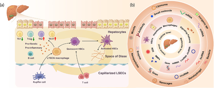
FIGURE 3.

(a) Schematic illustration of the pathogenesis of hepatic fibrogenesis. In chronic liver disease, damaged hepatocytes, Kupffer cells, and macrophages release chemokines and cytokines to activate hepatic stellate cells (HSCs), which produce excessive extracellular matrix. The activated HSCs aggravate hepatocyte injuries and induce capillarization and accumulation of liver sinusoidal endothelial cells. The proinflammatory and pro‐fibrotic factors released from the HSCs and immune cells create a positive feedback loop. (b) Summary of anti‐fibrotic targets at different biological scales. Anti‐fibrotic targets at molecular, cellular, and tissue levels and their corresponding therapeutic strategies are summarized based on the understanding of the mechanisms underlying intestinal fibrogenesis.
