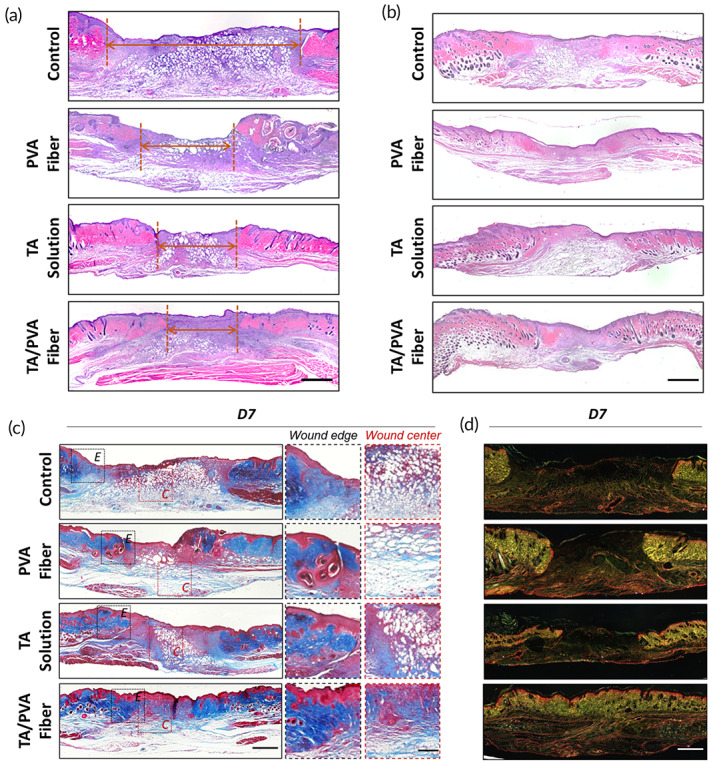
FIGURE 7.

Histological study of wound tissues. Hematoxylin and eosin (H&E) staining results of the wounds on Days 7 (a) and 14 (b). Red arrowheads represented the wound gaps. Scale bar = 1 mm. (c) Masson's trichrome staining result of wounds on Day 7. Both wound edge (E) and wound center (C) were enlarged for better observation. Scale bar for low magnification was 1 mm and 200 μm for high magnification. (d) Picrosirius red (PS) staining result of wounds on Day 7. Scale bar: 1 mm.
