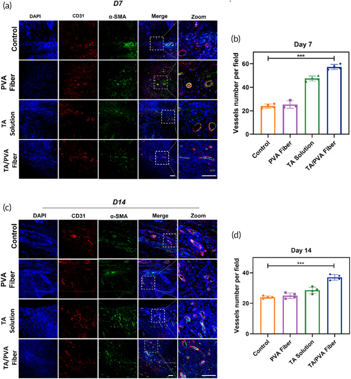
FIGURE 8.

Immunofluorescence staining of CD31 (red) and α‐smooth muscle actin (α‐SMA) (green) of wound tissues treated with PBS (Control), polyvinyl alcohol (PVA) fiber, tannic acid (TA) solution, and TA/PVA fiber on Days 7 (a) and 14 (c). The nuclei were stained as blue by 4′,6‐diamidino‐2‐phenylindole (DAPI). Statistical analysis of blood vessels on Days 7 (b) and 14 (d). Scale bars: 50 μm. Data were given as mean ± SD, ***p < 0.001, n = 4.
