Fig. 2.
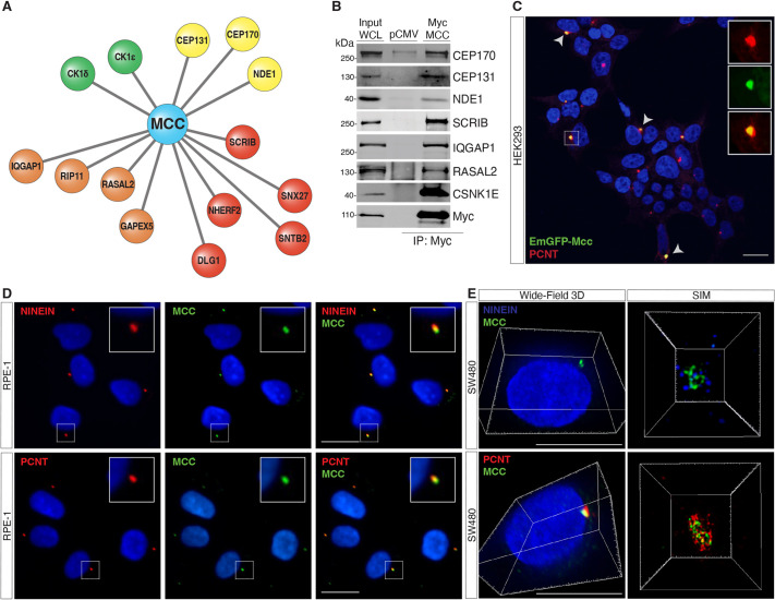
Proteomics analyses reveal MCC interactome. (A) Immunoprecipitation (IP) of FLAG-tagged human MCC in HEK293 cells followed by mass spectrometry identifies various MCC interactors, including centrosomal proteins (yellow), PDZ-domain-containing polarity proteins (red), GTPase regulators (orange), and kinases (CSNK1E/D) (green). (B) IP of Myc-tagged human MCC in HEK293 cells followed by western blotting for the indicated interactions. WCL, whole-cell lysate; pCMV, pCMV empty vector control. (C) HEK293 cells transfected with EmGFP–Mcc. Arrowheads indicate colocalization of EmGFP–Mcc with the centrosomal protein pericentrin (PCNT). Insets show single channels for EmGFP–Mcc and PCNT immunostaining. Scale bar: 20 μm. (D) Immunofluorescence (IF) for MCC and ninein or PCNT shows colocalization at the centrosome in human RPE-1 cells. (E) IF 3D widefield showing MCC colocalizing with PCNT and NINEIN. Scale bars: 50 μm. Super-resolution structured illumination microscopy (SIM) shows spatial proximity between MCC and ninein or PCNT at the centrosome in SW480 cells. All images shown are representative of at least five experiments.

