Fig. 3.
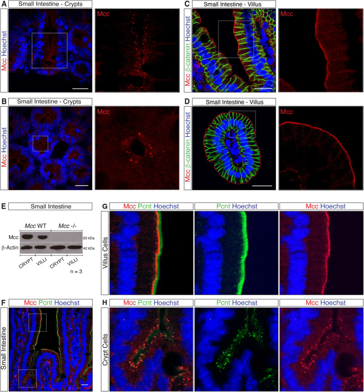
Mcc localizes to the centrosome in crypt cells and apical membrane of villus cells. (A–D) Immunofluorescence (IF) for Mcc in the mouse small intestine (SI). White-dashed squares highlight regions selected for higher magnification (A and C, longitudinal and B and D, transverse sections). (A,B) Punctate centrosomal staining for Mcc is observed in crypt cells. (C,D) IF for Mcc and β-catenin in SI villi. Mcc localizes to the apical membrane whereas β-catenin labels the lateral membrane of villus cells. Scale bars: 20 μm. (E) Western blot for Mcc in whole-cell lysates from purified crypt and villus fractions of the mouse SI. (F) IF showing colocalization of Mcc with the centrosomal protein Pericentrin (Pcnt) in the crypt and villus units of the mouse SI. White-dashed squares highlight regions selected for higher magnification shown in G and H. Scale bars: 20 μm. All images shown are representative of at least five experiments.

