
Fig. 3. Endothelial cells in BMoaC expressed proteins found in native bone marrow.
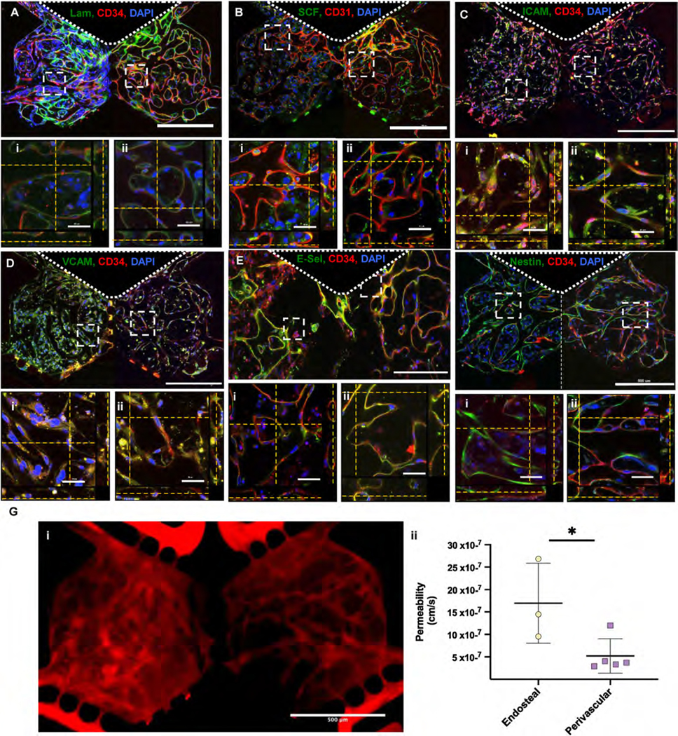
(A)–(F) Endothelial cell (red, CD31 or CD34) (A) laminin in the basement membrane, (B) SCF, (C) I-CAM, (D) V-CAM, (E) E-Selectin/CD62E and (F) Nestin. ROIs show z-stacks in either the (i) endosteal or (ii) perivascular niche. Nuclei were counterstained with DAPI (Blue). Scale bars: 500 μm and 50 μm for low and high magnification views, respectively. (G) BMoaC microvascular networks were i) perfused with 70 kDA TRITC-dextran and ii) had permeabilities of 16.96 × 10⁻⁷ cm/s and 5.19 × 10⁻⁷ cm/s for the endosteal (n = 3) and perivascular niches (n = 5), respectively.