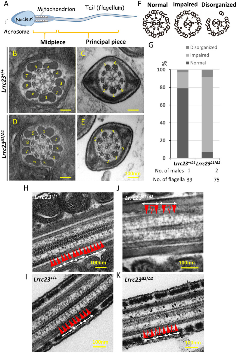
Fig. 5.
Ultrastructural analysis of cauda epididymis spermatozoa from Lrrc23Δ1/Δ1 mice. (A) An overview of the structure of a mature spermatozoon. (B-E) Electron microscopy images demonstrating normal radial spoke structures in wild-type spermatozoa (B,C) and radial spokes that were partially formed or absent in Lrrc23Δ1/Δ1 sperm (D,E). Outer dense fibers are marked with numbers. (F) Schematic representation of three axonemal structures of spermatozoa. (G) Percentages of the different axonemal structures in spermatozoa from Lrrc23+/+ and Lrrc23Δ1/Δ1 mice. (H-K) Longitudinal sections of the axonemal structures in spermatozoa from Lrrc23+/+ and Lrrc23Δ2/Δ2 mice. Radial spokes (red arrowheads, RS1-3) appeared in a repetitive pattern (average 96 nm repeat) in control (H,I). In Lrrc23 KO sperm flagella (J,K), the radial spokes were rather irregular and/or incomplete (unfilled red arrowheads) and missing.

