Fig. 5.
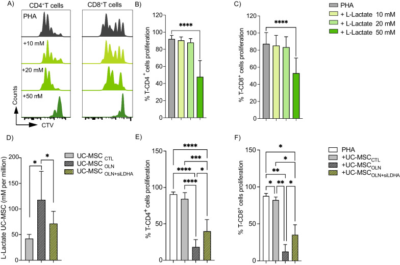
Lactate produced by UC-MSCs plays a critical role in their suppressive function. A–C PBMC were labeled with CTV and cultured alone or with 10, 20 or 50 mM L-lactate. After 4 days of culture, CD4+ (B) and CD8+ (C) T cells proliferation was analyzed by FACS. D Colorimetric analysis of lactate efflux by UC-MSCs after 24 h of treatment. E, F PBMCs labeled with CTV were cultivated alone (white bars) or co-culture with UC-MSCCTL (gray bars), UC-MSCOLN (striped, gray bars), or UC-MSCOLN+siLDHA (striped, yellow bars). Proliferation of CD4+ (E) and CD8+ (F) T cells was assessed by flow cytometry. Results represent the mean ± SD of three independent experiments for lactate measurement in UC-MSCs and two independent experiments with at least six different biological samples for PBMCs; *p < 0.05, **p < 0.01, ***p < 0.001, ****p < 0.0001 (unpaired Kruskal–Wallis test). Unless otherwise indicated, comparisons were with UC-MSCs in basal or control conditions or activated PBMCs

