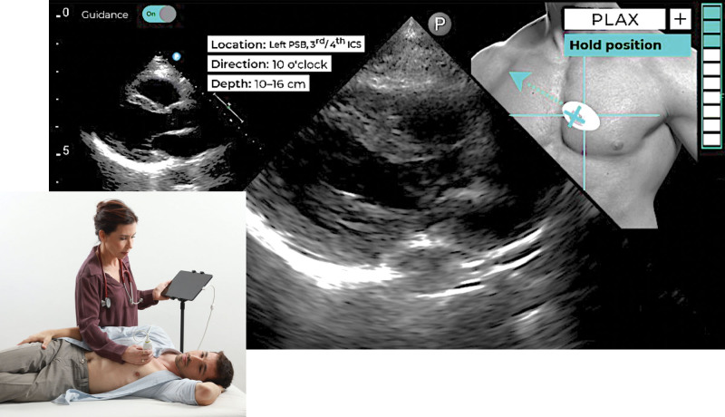
Figure 1.
Artificial intelligence–based echocardiographic image acquisition guidance system setup. An example of a system screen (right) showing the actual real-time view (middle) and visually intuitive instructions aimed at guiding the novice user how to manipulate transducer position and orientation toward achieving the target view (top, right), along with the ideal target view shown as a reference (top, left; see text for details). The quality bar is located on the top right corner of the screen. PLAX indicates parasternal long axis.

