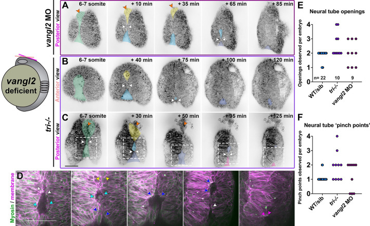
Figure 2. The neural folds of vangl2 deficient embryos exhibit ectopic closure points.
A-C) Still frames from time-lapse series of anterior neural tube development in vangl2 morphant (A) or tri−/− embryos (B-C) expressing membrane GFP or mCherry beginning at the 5-somite stage, viewed dorsally from more anterior (B) or posterior (A, C) positions. Green shading indicates the early neural groove, white arrowheads indicate pinch points at which the bilateral neural folds make contact. Yellow and blue shading indicate the initial anterior and posterior openings, respectively. Indigo and pink shading indicate new openings formed by “pinching” of the initial posterior opening. Each image series is a single Z plane from a confocal stack and is representative of 8 morphant and 10 mutant embryos imaged in 2 or 4 independent trials, respectively. D) Enlargements of regions in (C) within dashed boxes showing membrane Cherry in magenta and the Sf9-mNeon Myosin reporter in green. Arrows highlight Myosin localization to the edges of neural fold openings. The colors of the arrowheads correspond to the color of shading in (C). E-F) Quantification of the number of neural fold openings (E) and pinch points (F) observed in time-lapse series of embryos of the conditions indicated. Each dot represents a single embryo from 2 WT, 2 vangl2 morphant, and 4 tri independent trials. Anterior is up in all images, scale bars = 100 μm. See also Supp. videos 6–8.

