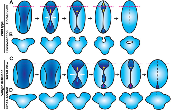
Figure 7. Model for anterior neural tube closure in zebrafish embryos.
A) Diagram of the anterior (brain region) neural plate in WT zebrafish embryos from approximately 4–10 somite stage, viewed from the dorsal surface with anterior to the top. A shallow neural groove (dark blue) forms at the dorsal midline between the bilateral neural folds (light blue). The neural folds come together at a central “pinch point” (white arrows), creating anterior and posterior openings. The posterior opening zippers closed caudally from the pinch point (cyan arrows) and the anterior opening goes on to form an eye-shaped opening in the forebrain region. The anterior edge of the eye-shaped opening zippers toward the posterior while its posterior edge zippers anteriorly, closing the eye-shaped opening from both sides (yellow arrows). The posterior opening continues to zipper toward the hindbrain until the neural folds in the entire brain region have fused. Dashed magenta lines represent the positions of the cross-sectional views shown in (B). B) Cross-sectional views of the anterior WT neural plate at the position of the dashed magenta lines in (A), dorsal is up. A U-shaped neural groove forms between the bilateral neural folds, which approach the midline and then fuse dorsally to enclose a hollow lumen. C) Diagram of anterior neural plate morphogenesis in vangl2 deficient embryos. The neural plate and groove begin wider and are delayed in the formation of the first pinch point (white arrows) and neural fold fusion. Additional pinch points form int the posterior opening, creating an additional opening that zippers closed (magenta arrows). D) Cross-sectional views of the anterior vangl2 deficient neural plate at the position of the dashed magenta lines in (C), dorsal is up. The forebrain forms a V-shaped neural groove that seals up from ventral to dorsal rather than enclosing a lumen.

