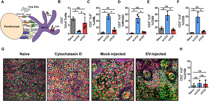
Figure 2: Tick EVs alter epidermal immune surveillance.
(A) Schematic representation of the DETC-keratinocyte crosstalk at the skin epidermis. (B-F, H) I. scapularis scV33 (blue) or siV33 (red) ticks were placed on C57BL/6 mice and allowed to feed for 3 days. On day 3, biopsies were taken from the skin at the bite site and compared to the naïve treatment (gray). (B) DETC (Vγ5), (C) JAML, (D) NKG2D, (E) CD69, (F) CD25, and (H) CD100 cells were assessed by flow cytometry. Graphs represent 1 of 3 independent experiments. (G) Epidermis containing Langerhans cells (red), DETCs (green), and keratinocytes (white) imaged on day 3 after injection with phosphate buffered saline (PBS - mock) or EV (4×10⁷ particles) into the mouse ear. Cytochalasin D (100 μg) was applied topically on the mouse ear every 24 hours for 2 days to induce DETC rounding as a positive control. Langerhans cells, DETCs and epithelial cells were simultaneously visualized in the huLangerin-CreER; Rosa-stop-tdTomato; CX3CR1-GFP+/−; K14-H2B-Cerulean mouse strain. Cre expression was induced with an intraperitoneal injection of tamoxifen (2 mg). Images from one out of three independent experiments. Statistical significance shown as *p<0.05, ns = not significant. Data are presented as a mean with standard deviation. Significance was measured by One-way ANOVA followed by Tukey’s post hoc test.

