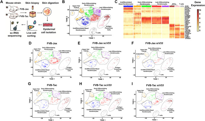
Figure 3: Epidermally-enriched scRNA-seq of the tick bite site.
(A) Overview of the experimental design. ScV33 and siV33 I. scapularis nymphs were placed on FVB-Jackson (FVB-Jax) or FVB-Taconic (FVB-Tac) mice and fed for 3 days. Skin biopsies at the bite site were digested with dispase and collagenase for epidermal cell isolation. Cells were sorted and prepared for scRNA-seq. (B) Composite tSNE plot of keratinocyte, T cell and antigen presenting cells in FVB-Jax and FVB-Tac mice in the presence or absence of I. scapularis nymphs microinjected with scV33 or siV33. tSNE plot represents 5,172 total cells following filtration as described in the materials and methods. (C) Heatmap depicting expression of the top 5 marker genes present in clusters from the epidermally enriched tSNE plot clusters (as shown in B). (D-I) Individual tSNE plots separated by mouse strain (FVB-Jax or FVB-Tac) in the presence or absence of I. scapularis nymphs microinjected with scV33 or siV33.

