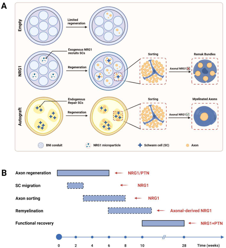
Figure 12. Schematic of cellular and molecular mechanisms involved in autograft and BNI critical gap repair.
A, Axon sorting and remyelination outcomes resulted from different treatment strategies. In an empty BNI conduit, the number of Schwann cells in the middle of the conduit was limited and axon regeneration mostly fails. NRG1 delivery recruits pro-regenerative SCs to migrate and proliferate in the conduit. However, due to a lack of axonal NRG1 type III expression, remyelination of the regenerated axons is limited. In an autograft treated animal, axon regeneration and remyelination was well supported by the repair Schwann cells. B, Timing of critical steps involved in nerve regeneration and the molecules regulating these steps. Dashed boxes are used to indicate the putative timing of the event.

