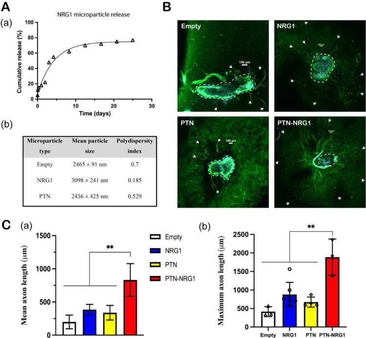
Figure 2. Microparticle release of NRG1.
A, (a) 28-day NRG1 release profile; (b) Characterization of the PLGA microparticle size and polydispersity index. B, Representative confocal images of DRG explants. DRGs were immunostained for axon (β-tubulin, green) and cell nuclei (DAPI, blue). Neurites were highlighted by white arrows while cell bodies were highlighted in yellow dashed circles. Scale bar: 100 μm. C, Quantification of the mean and maximum neurite length of DRG explants cocultured with empty, NRG1, PTN, and PTN-NRG1 microspheres for one week. **, p<0.01.

