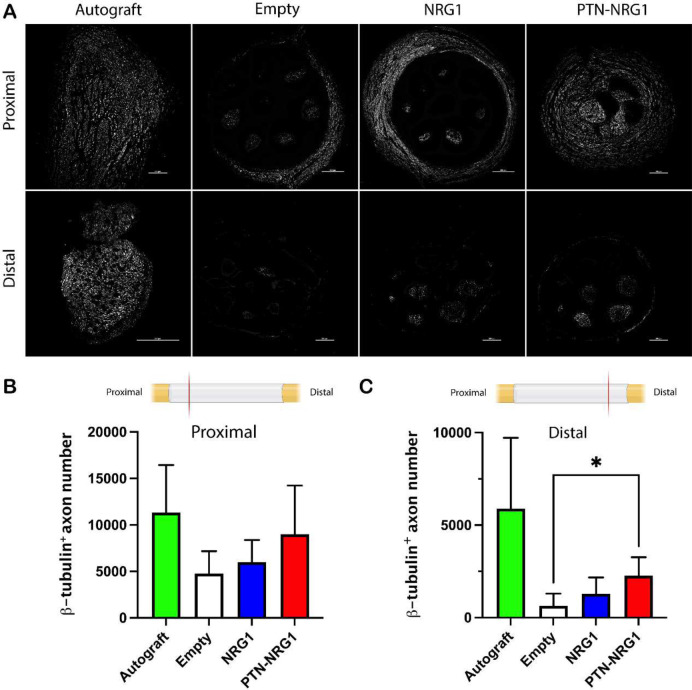
Figure 4. Axon regeneration into the distal nerve stump is limited.
A, Representative confocal images of β-tubulin III labeled axons showed limited regeneration into the distal segments. B-C, Quantification of the β-tubulin+ axon in the proximal (B) and middle (C) segments showed enhanced axon number in the PTN-NRG1 treated group but reduced compared to autograft repair. Scale bar: 200 μm. *, p<0.05.

