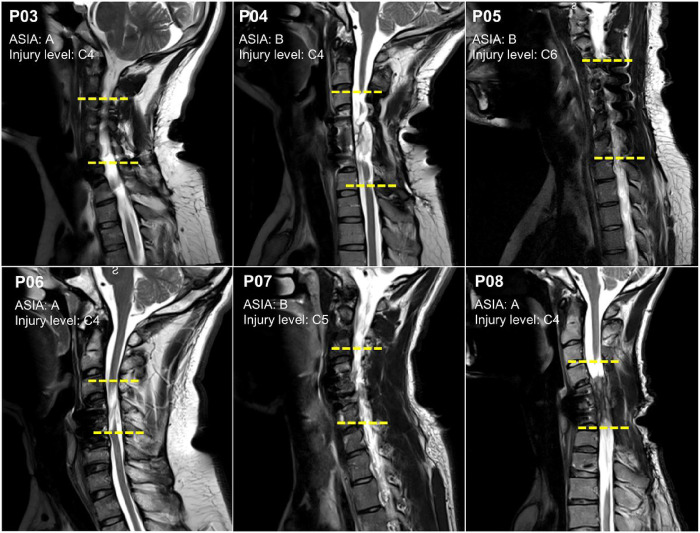
Figure 1. Structural magnetic resonance imaging (MRI) scans depicting sagittal sections within the cervical region.
Sagittal slices from six participants with cervical spinal cord injury classified as AIS A and B. The yellow horizontal lines indicate the location of the cervical lesion between the upper and lower vertebrae.

