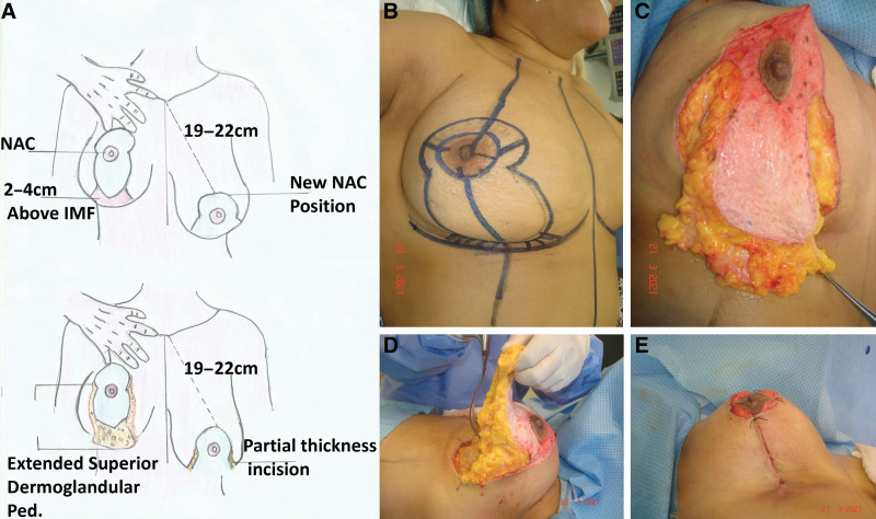
Fig. 1.
Diagram showing establishment of extended superior dermoglandular pedicle. A, Drawing, elevation and dissection of the extended superior pedicle starting from the IMF over the pectoral fascia. B, Drawing. C, Elevation of the extended portion from the inframammary area. D, Dissection of the extended pedicle from the pectoral fascia until just above the level of the nipple leaving the medial and lateral pillars. E, Shape of the breast after folding the extended portion 180 degrees over itself in the prepectoral pocket in cases of autoaugmentation mastopexy.

