
FIGURE 4.
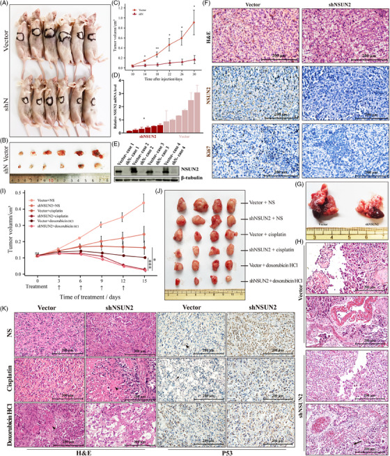
NSUN2 knockdown slows ATC growth in vivo and lung metastasis, increasing the efficacy of chemotherapy drugs. (A and B) Overview of tumours in xenograft mice model subcutaneously implanted with shN and vector cells. Scale bars, 200 μm. (C) Growth curves of tumour volumes formed by shN and vector cells. Data were presented as the mean ± SEM. (D) qRT‐PCR analysis of NSUN2 mRNA level in xenograft tumours. (E) WB showing the protein level of NSUN2 in xenograft tumours. (F) Haematoxylin–eosin staining and IHC staining of NSUN2 and Ki67 in tumour samples in xenograft mice model subcutaneously implanted with shN and vector cells. Scale bars, 200 μm. (G) Representative images of tumours in lung metastasis mice model intracardiac‐injected with shN and vector cells. shN cells injection developed less metastatic nodules than vector cells injection. (H) Haematoxylin–eosin staining of lung‐metastatic nodules in model intracardiac‐injected with shN and vector cells. Scale bars, 200 μm. (I) Overview of cisplatin‐ or doxorubicin HCl‐treated tumours in xenograft mice model subcutaneously implanted with shN and vector cells. NS indicates normal saline, as control. (J) Growth curves of cisplatin‐ or doxorubicin HCl‐treated tumour volumes formed by shN and vector cells. Data were presented as the mean ± SEM. (K) Haematoxylin–eosin and IHC staining of drug‐treated tumours in xenograft mice model subcutaneously implanted with shN and vector cells. Scale bars, 200 μm.