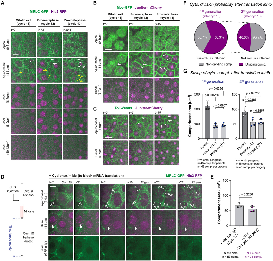
Figure 2. Cytoplasmic division cycles can occur without the nucleus or mRNA translation.

(A–C) Micrographs depict a rare fraction of cytoplasmic compartments (3.1% ± 2.1%) that can divide without nuclei (white and/or yellow arrows). A deeper image series is provided in (A) to demonstrate that there are no lurking nuclei.
(D) Translation inhibition by cycloheximide (CHX) does not prevent further cytoplasmic divisions (apico-basal), despite an arrest of nuclear cycle 10 (basal). Arrows and arrowheads highlight the dividing compartments and arrested nuclei, respectively. Images are a representative set from 4 embryos injected with the drug.
(E) Bar graphs for cytoplasmic compartment size under vehicle (H2O) or CHX conditions.
(F) Pie charts for the proportion of cytoplasmic compartments that can continue their divisions in the first and second generation after CHX injection.
(G) Bar graphs for maximum compartment sizes under CHX condition, in comparison with their daughter compartment sizes at the beginning of the next cycle. Analyses and experiments (E)–(G) were performed in embryos expressing MRLC-GFP and His2-RFP. Data (E) and (G) are represented mean ± SD, where each data point represents the average from each embryo. Statistical significance was assessed using a Welch’s t test (for Gaussian distributed data) or a Mann-Whitney test. Scale bars, 10 μm.
See also Figures S2 and S3.
