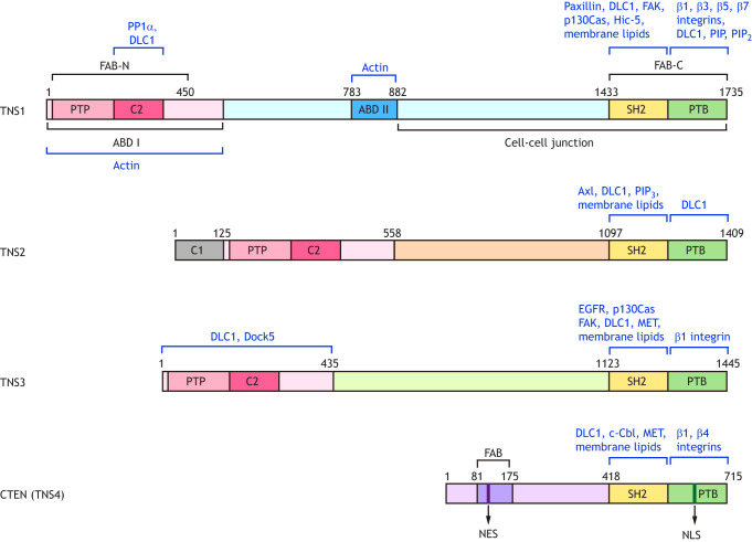
Fig. 1.
Domain structures of human tensins and their binding partners. The domains of tensins are represented by colored rectangles. The N-terminal regions of TNS1, TNS2 and TNS3 contain the actin-binding domain I (ABD I) that overlaps with the focal-adhesion-binding (FAB-N) site, as well as a PTEN-like protein tyrosine phosphatase (PTP) and C2 domains. The C-terminal regions of all tensins share the Src homology 2 (SH2) and phosphotyrosine-binding (PTB) domains that possess the FAB activity (FAB-C). The ABD II and the sequences required for proper cell–cell junction localization are unique to TNS1. The protein kinase C conserved region 1 (C1) is only present in TNS2. The N-terminal of CTEN (TNS4) contains a unique FAB domain, which includes a nuclear export sequence (NES), whereas a nuclear localization sequence (NLS) is located within the PTB domain. The binding partners of tensins mentioned in the main text are indicated in blue.

