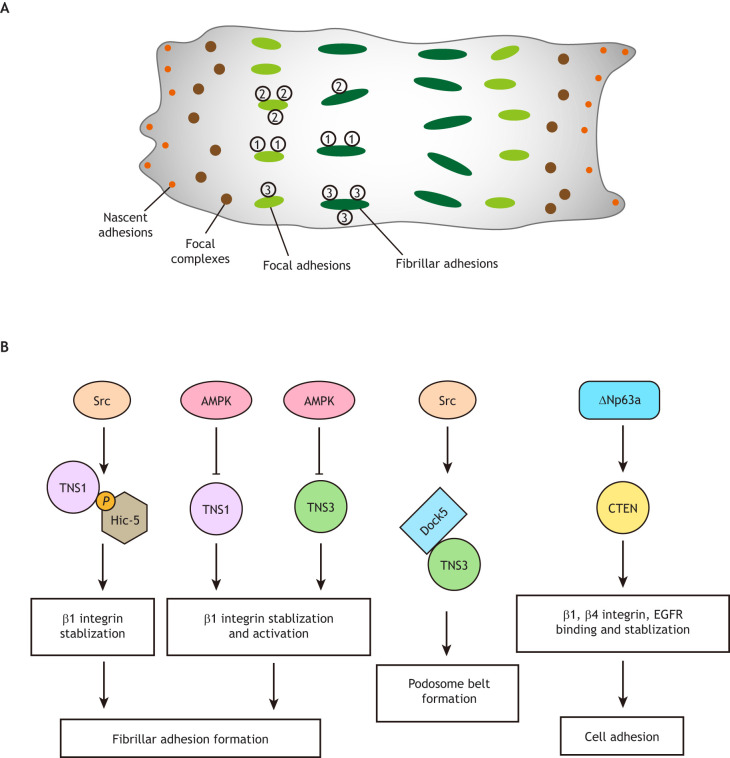
Fig. 2.
Cell adhesion structures and roles of tensins in cell adhesion. (A) Schematic representation of different subtypes of cell adhesion structures. Adherent cells initially form small nascent adhesions (orange dots), which develop into dot-like focal complexes (brown dots). Focal complexes progressively grow in size and mature into focal adhesions (green ovals), which then transform into elongated fibrillar adhesions (dark green oval). TNS1 (1), TNS2 (2), and TNS3 (3) are found in both focal and fibrillar adhesions. TNS2 is localized mainly in focal adhesions, TNS3 is mostly found in fibrillar adhesions, and TNS1 is distributed in both. (B) Tensins are required for cell adhesion. Src-dependent phosphorylation of Hic-5 interacts with TNS1 to promote β1 integrin stability and fibrillar adhesion maturation. AMP-activated protein kinase (AMPK) negatively regulates TNS1- and TNS3-dependent β1 integrin stabilization and activation, which is critical for fibrillar adhesion formation. TNS3 promotes podosome belt formation through a Src-dependent interaction with Dock5 in osteoclasts. CTEN expression is positively regulated by ΔNp63α, a transcription factor, and promotes cell adhesion through stabilizing β1 integrin, β4 integrin and EGFR.

