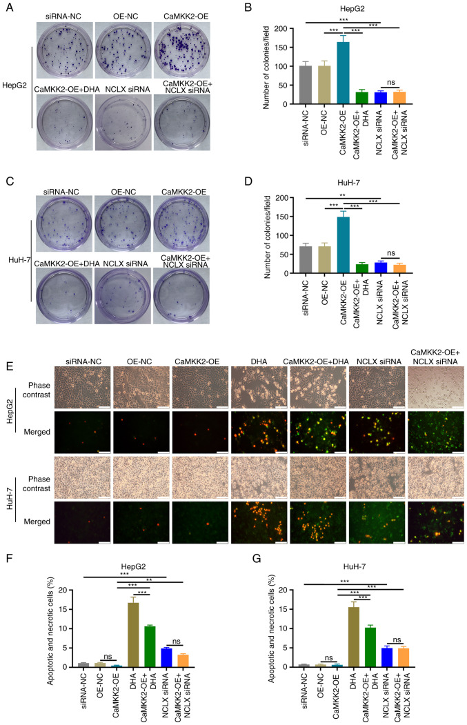
Figure 2.
Effects of DHA, CaMKK2 and NCLX on the proliferation and apoptosis of liver cancer cells. (A) Colony formation assay and (B) number of HepG2 colonies formed with cells treated with DHA, CaMKK2-OE and NCLX siRNA. (C) Colony formation assay and (D) number of HuH-7 colonies formed with cells treated with DHA, CaMKK2-OE and NCLX siRNA. Transfected (E) HepG2 and HuH-7 cells stained with YO-PRO-1 and PI dye and treated with DHA, CaMKK2-OE and NCLX siRNA. Apoptosis and necrosis rate of (F) HepG2 and (G) HuH-7 cells treated with DHA, CaMKK2-OE and NCLX siRNA. All data are presented as the mean ± standard deviation (n=3). Data were analyzed using one-way analysis of variance followed by Bonferroni post hoc test. Scale bar, 50 µm **P<0.01; ***P<0.001. ns, not significant; NCLX, mitochondrial sodium/calcium exchanger protein; DHA, dihydroartemisinin; CaMKK2, calcium/calmodulin-dependent protein kinase kinase 2; siRNA, small interfering RNA; OE, overexpression; NC, negative control.

