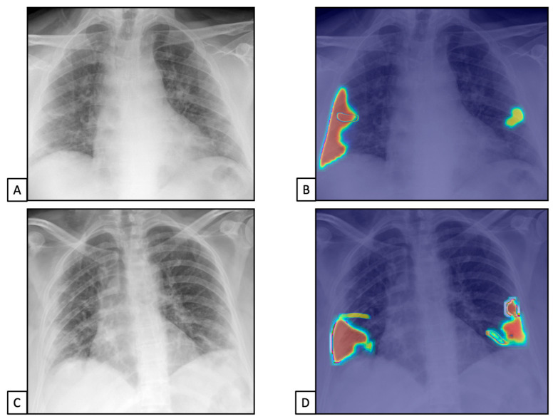
Figure 3.
Chest X-rays of patients admitted to the emergency department with the suspicion of COVID-19 infection belonged to dataset 1. (A,C) represent CXRs acquired at the bedside, showing multiple slight interstitial and alveolar opacities located peripherally and with a lower distribution. (B,D) The AI system analysis obtained in a few seconds displays the pathological zones. The AI system reported a high suspicion of COVID-19 infection (99.99%). The final diagnosis was lung involvement by COVID-19 pneumonia.

