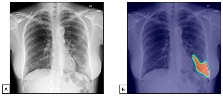
Figure 4.
Chest X-rays of patients admitted to the emergency department with respiratory distress and fever belonged to dataset 2. (A) represents CXR acquired at the bedside, showing compact typical opacities in the left lung lower zones. (B) represents AI system analysis, showing the pathological zones. The AI system reported a high suspicion of pneumonia, not typical for COVID-19 (99.99%). The final diagnosis was lung pneumonia due to Streptococcus pneumoniae.

