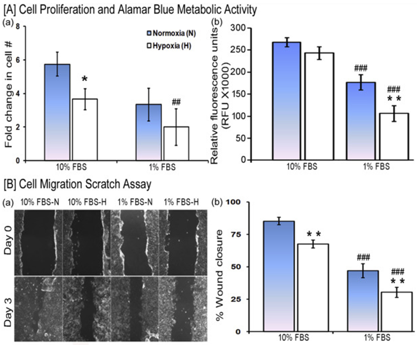
Fig. 1. Effect of hypoxia and low serum concentration on wound healing processes.

[A] HaCaT cell proliferation was evaluated after 3 days of culture in a medium supplemented with either 10% FBS or 1% FBS under normoxia and hypoxia. (a) Viable cell counts were relative to seeded cell number. (b) Metabolic activity estimated by the reduction of AlamarBlue reagent (n=9/each group). [B] Cell migration scratch assay. (a) Representative images of the scratch wound initially and after 3 days (10X). (b) Quantification of images comparing the fraction of the initial cell-free area in the scratch that has restored cell coverage. (n=6/each group). Statistical significance determined by ANOVA followed by post hoc Fisher's LSD test. * indicates a comparison between normoxia (N) and hypoxia (H) and # indicates a comparison between 10% FBS versus 1% FBS. */#P < 0.05, ##/**P < 0.01, and ###/***P < 0.001.
