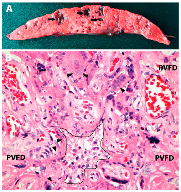
Figure 2.
Placental pathology in 3rd-trimester stillbirth attributed to SARS-CoV-2 placentitis. (A) Gross picture of a slice of placental tissue demonstrating diffuse effacement and consolidation of the parenchyma with scattered acute hemorrhagic cavities (arrows). (B) Microscopy showing features of SP, including mild mixed inflammatory infiltrate in the intervillous space (center, outlined), trophoblast necrosis and apoptosis (arrowheads), and perivillous fibrin deposition (PVFD). Original magnification ×400. Image from the author’s collection (the picture has not been previously published).

