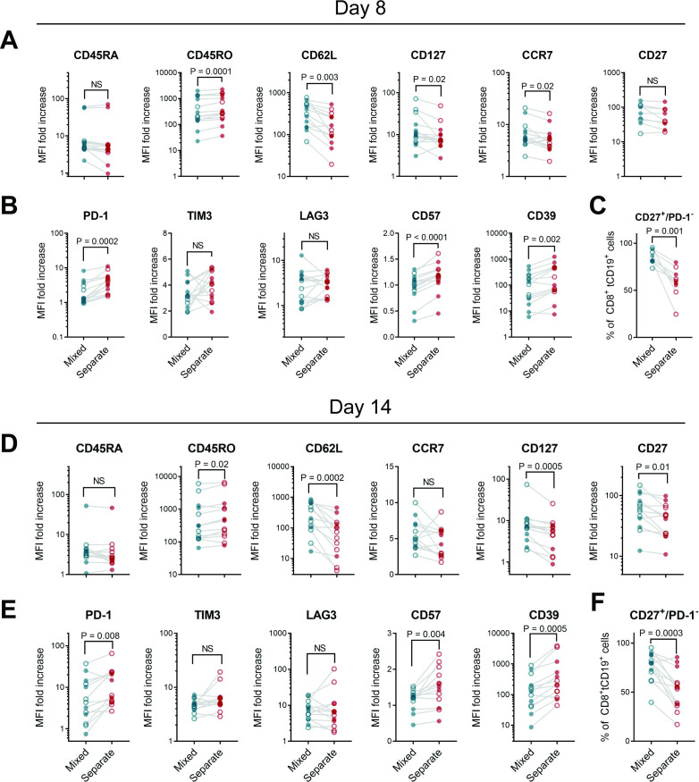
Figure 2.
Phenotypic differences between CD8+ CAR T cells cultured alone or those cultured with CD4+ T cells. CD4+ or CD8+ enriched PBMC from healthy donors (filled circles) or patients (open circles) were stimulated, then either mixed at a 60:40 CD4:CD8 ratio or maintained in separate cultures, then transduced with 1.5.3-NQ-28-BB-z CD20 CAR lentiviral vector. (A–C) Cells were harvested on day 8 of cell culture without restimulation, or (D–F) restimulated with irradiated CD20+ LCL cells on day 7 and harvested on day 14. Markers of memory and differentiation (A, D), exhaustion (B, E), or percentage of CD27+ PD1─ cells (C, F) were measured by flow cytometry. Gating strategy and representative histograms are shown in online supplemental figure 6. Data represent the fold increase in geometric mean fluorescence intensity (MFI) over isotype control, gated on viable CD8+ tCD19+ CAR T cells at day 7 (n=14: 5 patients and 9 healthy donors for A, B and n=9: 4 patients and 5 healthy donors for (C) or at day 14 (n=13: 7 patients and 6 healthy donors). P values were determined using paired two-tailed t-tests for markers meeting criteria for normality based on D’Agostino and Pearson or Shapiro-Wilk tests, or Wilcoxon matched-pairs signed rank test for markers not meeting normality criteria. CAR, chimeric antigen receptor; NS, not significant; PBMC, peripheral blood mononuclear cell.

