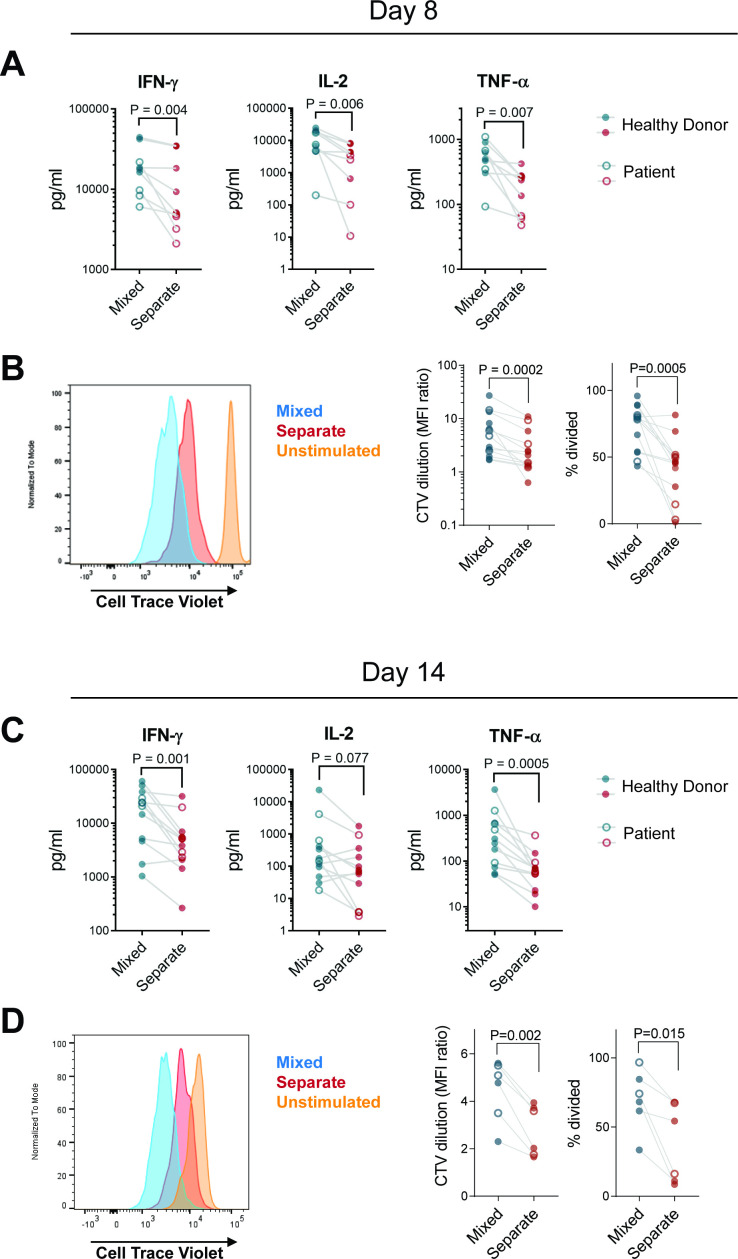
Figure 3.
CD8+ CAR T cells cultured in the absence of CD4+ cells have impaired cytokine secretion and proliferation in vitro. CD4+ and CD8+ enriched PBMC from patients (open circles) or healthy donors (filled circles) were stimulated, then either mixed at a 60:40 CD4:CD8 ratio or maintained in separate cultures, transduced with 1.5.3-NQ-28-BB-z CD20 CAR, and expanded. Cells were harvested on day 8 without restimulation (A, B) or restimulated with irradiated CD20+ LCL cells on day 7 and harvested on day 14 (C, D). FACS-sorted CD8+ tCD19+ T cells labeled with Cell Trace Violet (CTV) were incubated with irradiated CD20+ Raji-ffLuc cells (1:1 ratio). (A, C) supernatants were harvested at 24 hours and the indicated cytokines were measured by Luminex assay (n=9: 4 patients and 5 healthy donors for day 7; n=12: 4 patients and 8 healthy donors for day 14). (B, D) proliferation of the sorted cells after 4 days based on CTV dilution was assessed by flow cytometry. representative histograms are shown in the left panel, and summary data of geometric MFI ratio of unstimulated to stimulated cells and % divided cells are shown in the right panels (n=13: 4 patients and 9 healthy donors for day 7; n=6: 2 patients and 4 healthy donors for day 14). P values were determined using paired two-tailed t-tests for samples meeting criteria for normality based on D’Agostino and Pearson or Shapiro-Wilk normality test, or Wilcoxon matched-pairs signed RANK test for samples not meeting normality criteria. CAR, chimeric antigen receptor; MFI, mean fluorescence intensity; PBMC, peripheral blood mononuclear cell.

