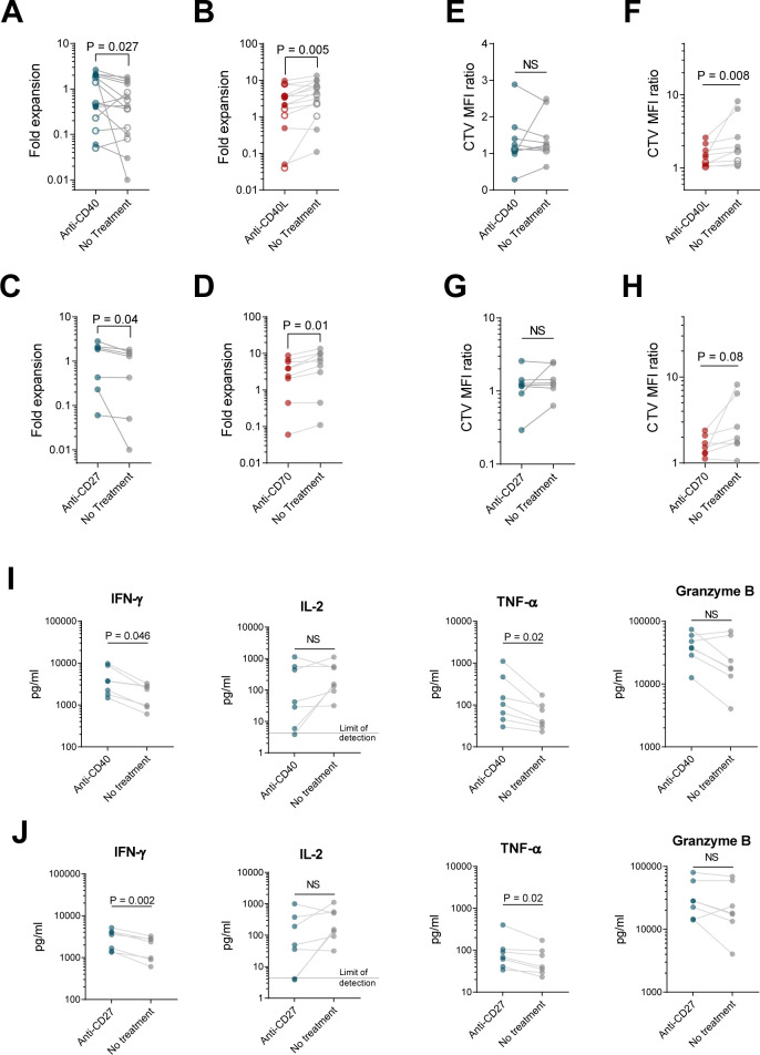
Figure 6.
CD4+ cells augment CD8+ T cell growth and function through both CD40L-CD40 and CD27-CD70 interactions. CD8+ cells from healthy donors (filled circles) or patients (open circles) were activated with αCD3/CD28 beads, cultured separately (A, C, E, G, I, J) or at a 60:40 ratio with CD4+ cells (B, D, F, H), transduced with 1.5.3-NQ-28-BB-z CD20 CAR lentivirus, and expanded in the presence of plate-bound agonistic anti-CD40 (A, E, I), plate-bound agonistic anti-CD27 antibody (C, G, J), soluble antagonistic anti-CD40L (B, F), or soluble antagonistic anti-CD70 antibody (D, H). (A–D) Fold expansion of CD8+ cells at day 8 is shown. (E–H) At day 8, cells were harvested, labeled with Cell Trace Violet (CTV), and restimulated with irradiated CD20+ Raji-ffLuc cells (1:1 ratio). Proliferation of the CD8+ cells after 4 days based on CTV dilution was measured by flow cytometry, with geometric MFI ratio of stimulated to unstimulated cells shown. (I–J) Supernatants from E, G were harvested 24 hours after restimulation, and levels of the indicated cytokines were measured by Luminex. Data represent mean values (±SEM). For (A), n=15 (6 patients and 9 healthy donors); for (B), n=14 (5 patients and 9 healthy donors); for (C), n=7 healthy donors; for (D), n=9 healthy donors; for (E, F, I), n=9 (2 patients and 7 healthy donors); for (G, H, J), n=7 healthy donors. P values were determined using paired two-tailed t-tests for samples meeting criteria for normality based on D’Agostino and Pearson or Shapiro-Wilk normality test, or Wilcoxon matched-pairs signed rank test for samples not meeting normality criteria. MFI, mean fluorescence intensity; NS, not significant.

