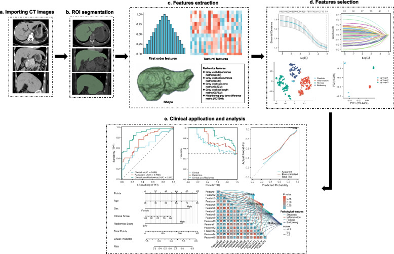
Fig. 4.
Evaluation of hepatic fibrosis based on radiomics. a Importing CT images. b The ROI was manually delineated on CT images of the entire liver. c First-order statistics, textural features, wavelet or Laplacian of Gaussian transforms, and shape features were extracted. d The feature selection is performed using a least absolute shrinkage, selection operator and cluster analysis, and cluster analysis, etc. e Nomogram was used to integrate radiomic and clinical features. The performance of established models was evaluated by receiver operator characteristic curve and precision-recall curve, the correlation between pathological features and radiomic features could be also analyzed, etc. CT Computed tomography, ROI Region of interest

