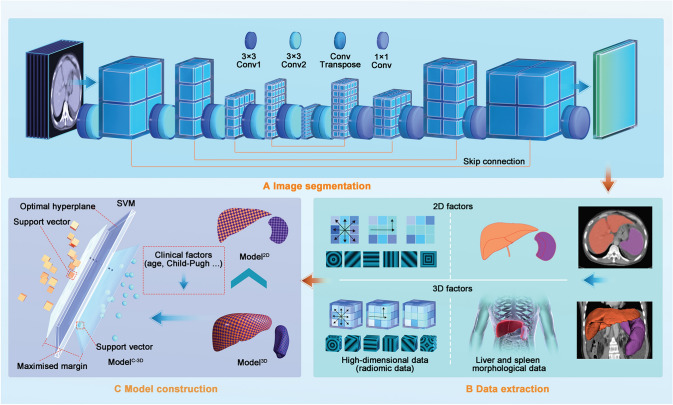
Fig. 2.
Workflow for model construction. Workflow for model construction. a Segmentation of liver and spleen on CT images using nnU-Net. b After extracting 2D and 3D factors (including morphologic and high-dimensional factors), we constructed 2D and 3D models. c Since the 3D model performed better than did the 2D model, we used the clinical and 3D factors to construct the combined model using the support vector machine. CT computed tomography

