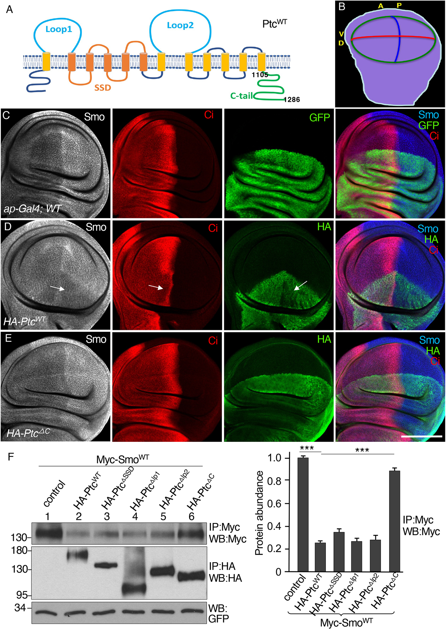
Fig. 1. The C-tail is required for Ptc to inhibit Smo.

(A) Ptc domain organization, with numbers indicating the amino acids positions of the C-tail. SSD, sterol-sensing domain. (B) A schematic drawing of a third instar larval wing disc, with anterior (A), posterior (P), dorsal (D), and ventral (V) compartments indicated. The blue line indicates the A/P boundary, the red line indicates the D/V boundary, and the green circle indicates the wing pouch region. (C) A wild-type (WT) wing disc expressing GFP under control of the dorsal compartment–specific ap-Gal4 driver and stained for Smo and Ci. N=3 wing discs. (D and E) Wing discs expressing HA-PtcWT (D) or HA-PtcΔC (E) under control of the ap-Gal4 driver stained for Smo, Ci and HA. Arrows in (D) indicate the region of the HA-PtcWT expression domain in the anterior compartment with decreased Smo, Ci, and HA-PtcWT. N=5 (D) or 4 (E) wing discs. (F) S2 cells were transfected with Myc-SmoWT in combination with the indicated HA-tagged Ptc variants. Identical volumes of cell lysates were immunoprecipitated (IP) and blotted (WB) with antibodies specific for Myc or HA. GFP served as transfection and lysate control. The density of each Myc-SmoWT band was quantified using ImageJ software. N=3 independent experiments. *** indicates a p < 0.001 (Student’s t test). Scale bar, 50 μm.
