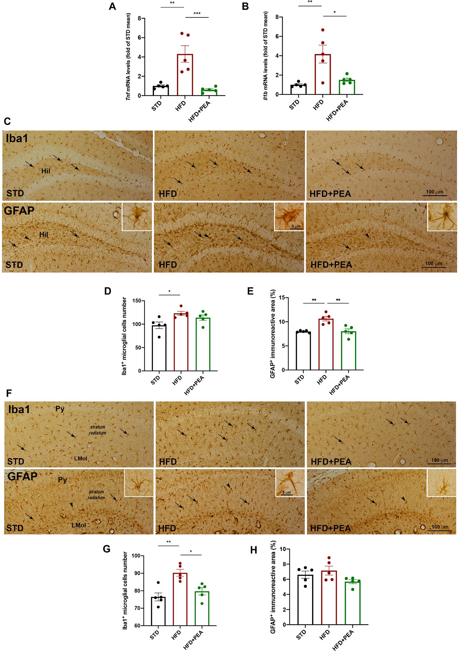
Fig. 5. PEA limits neuroinflammation, astrogliosis and microgliosis in the hippocampus of HFD mice.

The mRNA expression of (A) Tnf and (B) Il1b in the hippocampus of all experimental groups (n = 5 per group). The immunohistochemical analyses of Iba1-positive microglial cells and GFAP-positive astrocytes in (C) the DG and (F) stratum radiatum (n = 5 per group). The count of the number of positive cells for (D, G) Iba1 and (E, H) of GFAP immunoreactive in DG and stratum radiatum, respectively (n = 5 per group). Hil, hilus of the DG; Py, pyramidal cell layer; LMol, lacunosum molecular layer. All data are shown as mean ± S.E.M. *p < 0.05, **p < 0.01, and ***p < 0.001.
