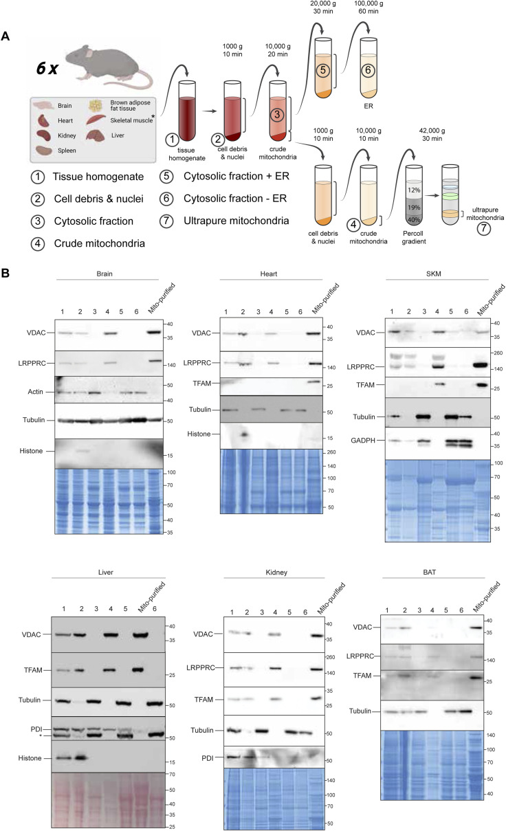
Figure S1. Validation of mitochondrial enrichment procedure by immunoblotting.
(A) Illustration of the isolation of the subcellular fractions of the different tissues. (B) Verification of purity of samples. Western blots were performed on small aliquots collected from subcellular fractions (1–6 and ultrapure mitochondria) that were isolated from different tissues (BAT, brain, heart, liver, kidney, and skeletal muscle, SKM), all from one mouse that is included in this study. Different antibodies against cellular markers were used to verify the purity of the different fractions: cytosol: actin/GADPH/tubulin; ER: PDI; mitochondria: VDAC (outer mitochondrial membrane), LRPPRC/TFAM (Matrix); nucleus: histone H3. Loading: Coomassie blue/Ponceau S stain. Bands of protein ladder (Thermo Fisher Scientific Spectra Multicolor Broad Range) in kD are illustrated on the right side. The asterisk marks the tubulin band from the former incubation. Note that no aliquot of purified mitochondria could be collected from spleen because all of the material was needed for the phosphoproteomic analysis.

