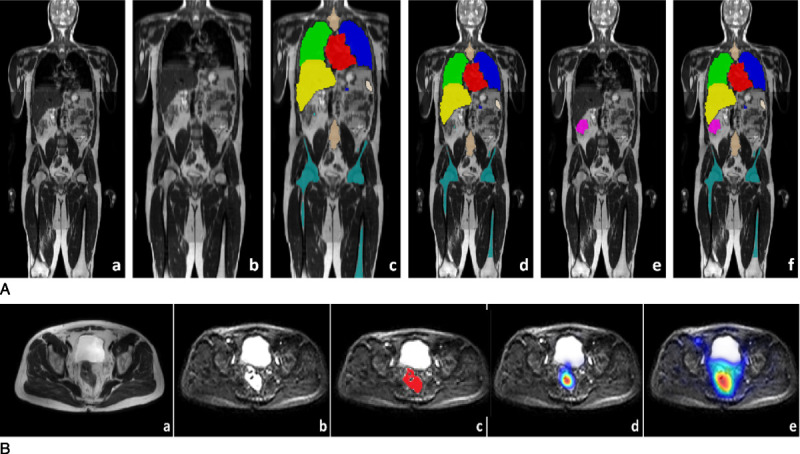
FIGURE 2.

Data generation process for the 2-stage model training approach. Panel A: A, An example of a T2WI WB-MRI scans from a participant in training set. B, After registration to a template scan from the healthy volunteer study. C, Output of the organ. Segmentation algorithm developed in healthy volunteer study. D, After mapping the organ segmentations back to the original scan from training data. E, Manual lesion segmentation overlaid on the T2WI scan. F, Merged organ segmentations and cancer lesion segmentation overlaid on the T2WI scan, which is used for training the final multiclass segmentation algorithm. Panel B: Cancer lesion detection training. A, Input T2WI scan (different patient to panel A). B, Diffusion-weighted scan. C, Manual lesion segmentation (based on reference standard) from T2WI image overlaid on diffusion scan. D, Postprocessed lesion probability map from the convolutional neural network (CNN) algorithm (deep medic). E, Postprocessed lesion probability map from the classification forest (CF) algorithm.
