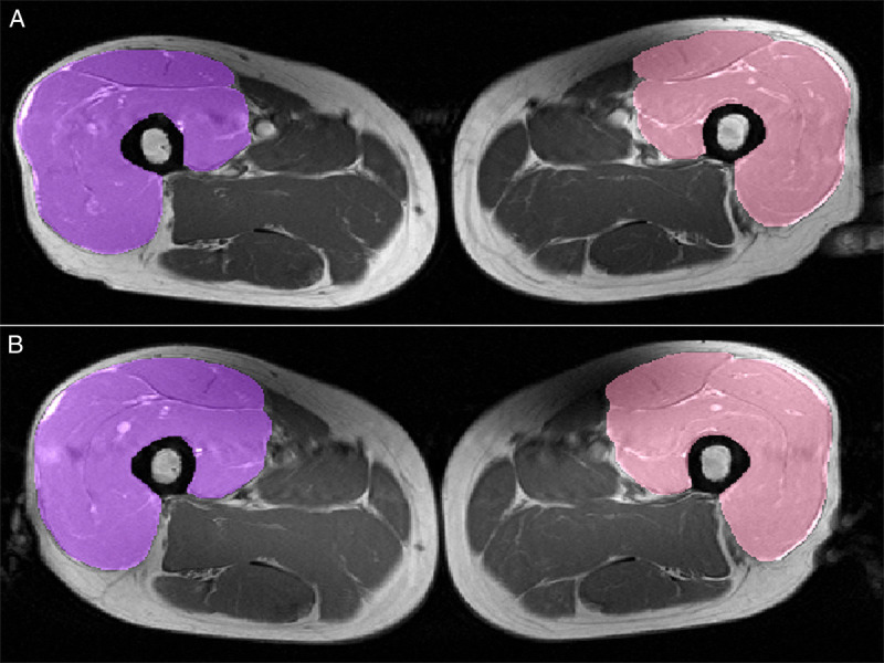
FIGURE 2.

Muscle segmentation in MR images before and after training intervention. T1-weighted MR images with segmentation of the quadriceps femoris muscle (patient’s right side in purple and left side in pink) at baseline (a; top) and after the exercise training intervention (b; bottom). The femur, fatty, and connective tissues as well as vessels were excluded from the segmentation, but fascia lata and beginning rectus tendon were included. This technique and the slice-selection have been shown to adequately represent changes in extensor muscle volume (r2 = 0.73) (34).
