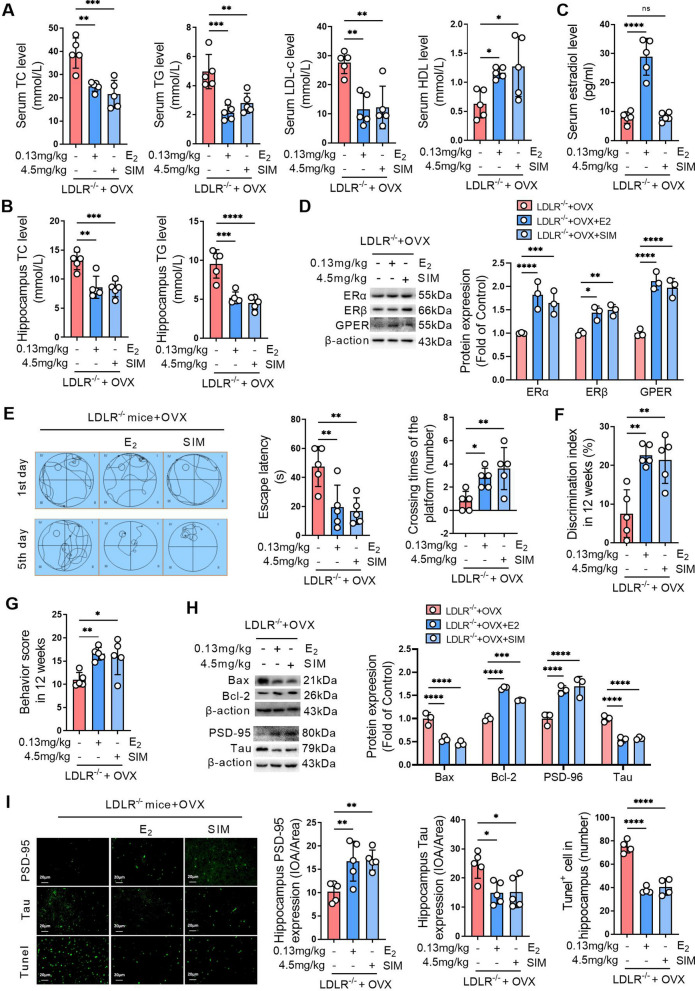
Fig. 7.
Activating ERs improves hippocampal damage and cognitive impairment caused by ovariectomy in LDLR−/− mice. Bilateral ovariectomy was performed in LDLR−/− mice fed a HFD to simulate a postmenopausal stage. The mice were treated with 0.13 mg/kg estradiol (E2) or 4.5 mg/kg simvastatin (SIM) for 90 days. A Serum of mice was obtained after 90 days of treatment, and the levels of TC, TG, LDL-c and HDL-c were detected using biochemical kits. B The levels of TC and TG in hippocampal homogenates were detected using biochemical kits. C The serum estradiol levels were detected using an ELISA kit. D The expression of ERα, ERβ and GPER in the hippocampal tissues of mice was detected by western blot, and the relative quantitative analysis of ERα, ERβ and GPER expression was performed. E The learning and memory ability of mice was evaluated by the Morris water maze after 90 days of treatment. The left panel shows the swimming paths of mice on the first and fifth training days of the Morris water maze test. The middle panel indicates the escape latency of mice on the fifth training day of the Morris water maze test. The right panel indicates the number of platform crossings on the sixth day of the Morris water maze test. F The learning and memory abilities of mice were evaluated by the novel object recognition test after 90 days of treatment. G Spatial memory of mice was evaluated by the Y-maze task after 90 days of treatment. H The expression of Bax, Bcl-2, PSD-95, and Tau in the hippocampal tissues of mice was detected by western blot, and the relative quantitative analysis of Bax, Bcl-2, PSD-95, and Tau expression was performed. I The expression of PSD-95 and Tau was detected by immunofluorescence staining, and the cell apoptosis was detected by tunel staining in the hippocampal tissues of mice after 90 days of administration. The magnification and scale bar are marked in the representative images. The expression levels of PSD-95 and Tau in the hippocampus were quantified by the mean optical density, and the number of apoptotic cells in each view was counted. A, B and E–G, n = 5. D and H, n = 3. I, n = 4–5. In the indicated comparison, *P < 0.05, **P < 0.01, ***P < 0.001, ****P < 0.0001

