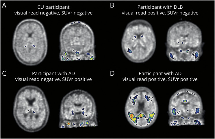
Figure 3. Example [18F]Flortaucipir PET Scans for Visual Read.
Shown are [18F]flortaucipir PET scans of 4 participants. (A) A CU participant defined as tau-negative on both visual read and SUVr. (B) A participant with DLB defined as visual read tau-positive, but SUVr negative. Increased tracer uptake was observed in only a small region, potentially resulting in low SUVr. (C) A participant with AD defined as visual read negative, but SUVr positive. Increase tracer uptake was observed isolated to the medial temporal lobe, which does not contribute to a positive tau-PET visual read. (D) A participant with AD defined as tau-positive on both visual read and SUVr. AD = Alzheimer disease; CU = cognitively unimpaired; DLB = dementia with Lewy bodies; SUVr = standardized uptake value ratio.

