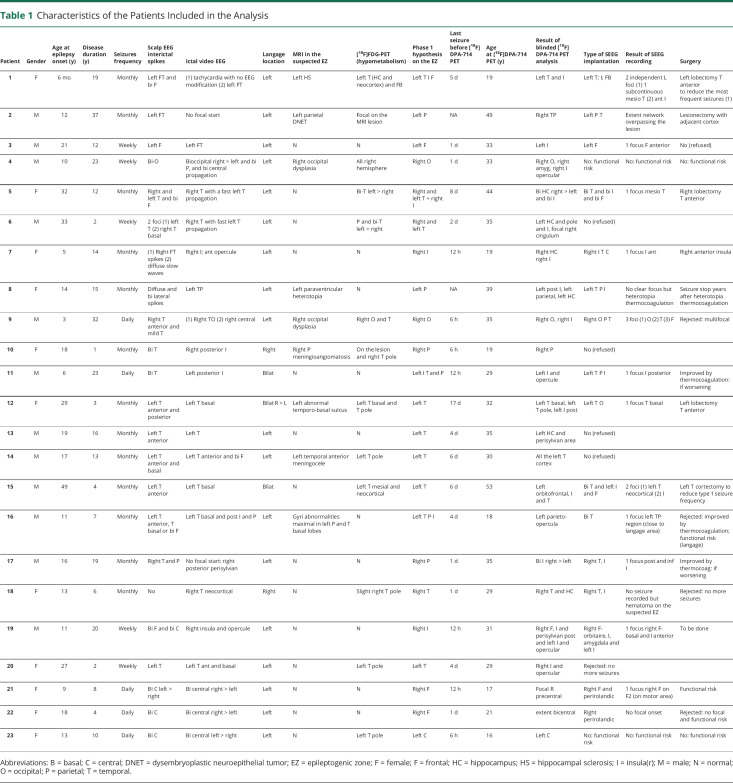
Table 1.
Characteristics of the Patients Included in the Analysis
| Patient | Gender | Age at epilepsy onset (y) | Disease duration (y) | Seizures frequency | Scalp EEG interictal spikes | Ictal video EEG | Langage location | MRI in the suspected EZ | [18F]FDG-PET (hypometabolism) | Phase 1 hypothesis on the EZ | Last seizure before [18F]DPA-714 PET | Age at [18F]DPA-714 PET (y) | Result of blinded [18F]DPA-714 PET analysis | Type of SEEG implantation | Result of SEEG recording | Surgery |
| 1 | F | 6 mo | 19 | Monthly | Left FT and bi F | (1) tachycardia with no EEG modification (2) left FT | Left | Left HS | Left T (HC and neocortex) and FB | Left T I F | 5 d | 19 | Left T and I | Left T; I; FB | 2 independent L foci (1) 1 subcontinuous mesio T (2) ant I | Left lobectomy T anterior to reduce the most frequent seizures (1) |
| 2 | M | 12 | 37 | Monthly | Left FT | No focal start | Left | Left parietal DNET | Focal on the MRI lesion | Left P | NA | 49 | Right TP | Left P T | Extent network overpassing the lesion | Lesionectomy with adjacent cortex |
| 3 | M | 21 | 12 | Weekly | Left F | Left FT | Left | N | N | Left F | 1 d | 33 | Left I | Left F | 1 focus F anterior | No (refused) |
| 4 | M | 10 | 23 | Weekly | Bi-O | Bioccipital right > left and bi P, and bi central propagation | Left | Right occipital dysplasia | All right hemisphere | Right O | 1 d | 33 | Right O, right amyg, right I opercular | No: functional risk | No: functional risk | No: functional risk |
| 5 | F | 32 | 12 | Monthly | Right and left T and bi F | Right T with a fast left T propagation | Left | N | Bi-T left > right | Right and left T + right I | 8 d | 44 | Bi HC right > left and bi I | Bi T and bi I and bi F | 1 focus mesio T | Right lobectomy T anterior |
| 6 | M | 33 | 2 | Weekly | 2 foci (1) left T (2) right T basal | Right T with fast left T propagation | Left | N | P and bi-T left > right | Right and left T | 2 d | 35 | Left HC and pole and I, focal right cingulum | No (refused) | ||
| 7 | F | 5 | 14 | Monthly | (1) Right FT spikes (2) diffuse slow waves | Right I; ant opercule | Left | N | N | Right I | 12 h | 19 | Right HC right I | Right I T C | 1 focus I ant | Right anterior insula |
| 8 | F | 14 | 15 | Monthly | Diffuse and bi lateral spikes | Left TP | Left | Left paraventricular heterotopia | N | Left P | NA | 39 | Left post I, left parietal, left HC | Left T P I | No clear focus but heterotopia thermocoagulation | Seizure stop years after heterotopia thermocoagulation |
| 9 | M | 3 | 32 | Daily | Right T anterior and mild T | (1) Right TO (2) right central | Left | Right occipital dysplasia | Right O and T | Right O | 6 h | 35 | Right O, right I | Right O P T | 3 foci (1) O (2) T (3) F | Rejected: multifocal |
| 10 | F | 18 | 1 | Monthly | Bi T | Right posterior I | Right | Right P meningioangiomatosis | On the lesion and right T pole | Right P | 6 h | 19 | Right P | No (refused) | ||
| 11 | M | 6 | 23 | Daily | Bi T | Left posterior I | Bilat | N | N | Left I T and P | 12 h | 29 | Left I and opercule | Left T P I | 1 focus I posterior | Improved by thermocoagulation: if worsening |
| 12 | F | 29 | 3 | Monthly | Left T anterior and posterior | Left T basal | Bilat R > L | Left abnormal temporo-basal sulcus | Left T basal and T pole | Left T | 17 d | 32 | Left T basal, left T pole, left I post | Left T O | 1 focus T basal | Left lobectomy T anterior |
| 13 | M | 19 | 16 | Monthly | Left T anterior | Left T | Left | N | N | Left T | 4 d | 35 | Left HC and perisylvian area | No (refused) | ||
| 14 | M | 17 | 13 | Monthly | Left T anterior and basal | Left T anterior and bi F | Left | Left temporal anterior meningocele | Left T pole | Left T | 6 d | 30 | All the left T cortex | No (refused) | ||
| 15 | M | 49 | 4 | Monthly | Left T anterior | Left T basal | Bilat | N | Left T mesial and neocortical | Left T | 6 d | 53 | Left orbitofrontal, I and T | Bi T and left I and F | 2 foci (1) left T neocortical (2) I | Left T cortectomy to reduce type 1 seizure frequency |
| 16 | M | 11 | 7 | Monthly | Left T anterior, T basal or bi F | Left T basal and post I and P | Left | Gyri abnormalities maximal in left P and T basal lobes | N | Left T P I | 4 d | 18 | Left parieto-opercula | Bi T | 1 focus left TP region (close to langage area) | Rejected: improved by thermocoagulation; functional risk (langage) |
| 17 | M | 16 | 19 | Monthly | Right T and P | No focal start: right posterior perisylvian | Left | N | N | Right P | 1 d | 35 | Bi I right > left | Right T, I | 1 focus post and inf I | Improved by thermocoag: if worsening |
| 18 | F | 13 | 6 | Monthly | No | Right T neocortical | Right | N | Slight right T pole | Right T | 1 d | 29 | Right T and HC | Right T, I | No seizure recorded but hematoma on the suspected EZ | Rejected: no more seizures |
| 19 | M | 11 | 20 | Weekly | Bi F and bi C | Right insula and opercule | Left | N | N | Right I | 12 h | 31 | Right F, I and perisylvian post and left I and opercular | Right F-orbitaire, I, amygdala and left I | 1 focus right F-basal and I anterior | To be done |
| 20 | F | 27 | 2 | Weekly | Left T | Left T ant and basal | Left | N | Left T pole | Left T | 4 d | 29 | Right I and opercular | Rejected: no more seizures | ||
| 21 | F | 9 | 8 | Daily | Bi C left > right | Bi central right > left | Left | N | N | Right F | 12 h | 17 | Focal R precentral | Right F and perirolandic | 1 fucus right F on F2 (on motor area) | Functional risk |
| 22 | F | 18 | 4 | Daily | Bi C | Bi central right > left | Left | N | N | Right F | 1 d | 21 | extent bicentral | Right perirolandic | No focal onset | Rejected: no focal and functional risk |
| 23 | F | 13 | 10 | Daily | Bi C | Bi central left > right | Left | N | Left T pole | Left C | 6 h | 16 | Left C | No: functional risk | No: functional risk | No: functional risk |
Abbreviations: B = basal; C = central; DNET = dysembryoplastic neuroepithelial tumor; EZ = epileptogenic zone; F = female; F = frontal; HC = hippocampus; HS = hippocampal sclerosis; I = insula(r); M = male; N = normal; O = occipital; P = parietal; T = temporal.

